Thursday, July 29, 2010

A New Classification of Palatal vaults: A Clinical Approach


I am presenting here a new classification for the Palatal vault in a completely edentulous patient.

Need for a classification: The general classification of palatal vaults as given by House is High, Medium and Low.
What does this indicate? It gives some idea about the depth of the palate, but so long as this information cannot be applied to a particular step in the fabrication of the denture, it has limited value! Should we do something different if a patient is categorised as House's High palate? or low?... In which area of the maxilla do we actually measure and assess that the vault is high or medium or low?

Therefore I have come up with a new classification that co relates the palatal vault to phonetic requirements.

In this classification the depth of the palate is measured at the 1st canine - premolar level, assuming that the tongue makes maximum contact in that zone of the palate during speech, especially linguo-palatal sounds.


Methodology employed :
After denture try-in, the region of the canine-1st premolar is marked on
 the ridge of the cast. An imaginary line joining these bilateral points is
 used as a reference and a perpendicular drawn from this line to the
 surface of the palate on the cast gives us the depth of the palate.


Based on this depth as measured in a large section of edentulous patients in our society, 3 categories of palatal vaults have been derived:


  SK1 - Shallow palatal vault. Depth : 0-5mm
SK2 - Medium palatal vault. Depth : 6-10mm 
                
SK3 - Deep palatal vault. Depth : greater than 11mm

This classification was derived from a study of measuring palatal depths by the above method for 50 patients.


Significance of the new classification:
Since it is based on the palatal depth in the region of the canine-1st premolar,(which is the most common region of the articulation of the tongue with the palate)it can be directly linked to phonetic requirements of complete dentures.
The canine-1st premolar region is the region where the palate can give an L beam effect to a denture. One would expect that SK3 category patients will have greater denture stability. The classification can thus give a prognosis of the denture stability.I am presently studying whether in patients with varying palatal depths, (Whether a patient is SK1 or SK2 or SK3 category); does the incorporation of palatal rugae make any difference to the speech of the patient or not?

Palatal Rugae and Speech in Completely Edentulous cases of varying palatal vault depth.


This project is ongoing. Presented here is a report of my Pilot study:
The role of palatal rugae in speech of complete denture patients has been debatable. While some authors have questioned their role in the past, a few authors have also said that rugae incorporated on the palatal surface of                        a denture enhance pronunciations.

I have found that going the extra mile with edentulous patients to give them better speech is worth it as:

  • It is our responsibility
  • It is often neglected and left for the patient to adjust to.
  • Patients often complain that rugae has been replaced by smooth surface resulting in improper articulation.
  • Psychological comfort.


What I basially did was derived my own new classification for palatal-vault depths and tried to assess whether the presence or absence of rugae affects the speech in patients with varying depths of the palate.

Objectives of my project:
To compare and evaluate pronunciation of linguo-palatal sounds in
patients with high, medium and low palatal vault depths using trial
record bases with and without palatal rugae.

Inclusion Criteria for my patients:

-Normal speech.
-Normal hearing.
-Good neuromuscular control ;voluntary control of tongue,lips and cheek.
-Read standard paragraph given for recital for speech assessment.
-Normal ridge relationship.
-Ridges without prominent undercuts.

Exclusion criteria for my patients:

-Speech abnormalities.
-Impaired hearing.
-Poor neuromuscular control.
-Inability to read.
-Undercuts which would needed to be blocked prior to fabrication.
-Previous denture wearers who have developed adjusted speech patterns.
-Unwilling/uncooperative patients.


Methodology:

Impressions were made using routine standard procedures.
Record bases were fabricated for each patient using a uniform thickness of autopolymerising acrylic resin.
Jaw relation recording and mounting on a Mean Value articulator was done.
Denture teeth were selected and set up on the rims.
The trial dentures were approved by the patient for their esthetic value.

Following this step the patients were divided in 3 categories (see separate blog for details):
SK1 - Shallow palatal vault
SK2 - Medium depth palatal vault
SK3 - Deep palatal vault.

Each patient underwent speech testing. Initial testing was done using maxillary and mandibular trial dentures with rugae incorporated with wax. Subsequently the rugae were eliminated and speech test was repeated using a smooth palatal contour.

Speech testing : Each patient was made to read a paragraph containing and stressing on 't', 'd', 'n', 'l' sounds.


Assessment was done in a sound proof room by 2 experienced speech pathologists and 1 lay person.
The testing procedure was double blinded as the assessors and the patients were not told about which set of record bases were being used (with or without the rugae).


1 patient was tested of each palatal vault depth.

The Speech Rating Scale used (Developed by Speech language Pathology Dept, AYJNIHH,1984)

0-Normal.

1-Can understand without difficulty. However feels speech is not normal.

2-Can understand with little efforts.

3-Can understand with concentration and effort, especially by a sympathetic listener.

4-Can understand with difficulty and concentration by family, but not by others.

5- Can understand with effort if context is known.

6-Cannot understand at all, even when context is known.


The results of the 3 patients can be summarised as follows


                         Without dentures       Dentures with rugae        Dentures without rugae
SK1(Shallow) -       1.6                                  2 .0                                     2.3

SK2(Medium) -       1.0                                  1.3                                      0.6

SK3 (Deep) -           0.6                                 1.0                                      1.3

The shallow and deep palate showed an improvement in phonetics when rugae was incorporated.
More studies and more patients need to be assessed to reach a definite conclusion


The project is ongoing and will be updated when I finish 10 patients in each SK category.

Friday, July 23, 2010

Belfast - Sept 09 IMPT Conference


The Institute of Maxillofacial Prosthetists and Technologists (IMPT) held their biannual conference at Belfast in September 2009. I was thrilled to be able to go to this Congress and read my research paper.

This conference is organised by the IMPT, UK and their website can be accessed at www.impt.org


It was a scary thought to go and lecture to a bunch of specialists about their own speciality, but they all put me at ease.

I presented my work on the Hardness changes in Maxillofacial elastomers.

    The conference was vastly different from the ones I have attended in India. Different in terms of its punctuality, organisation and execution. No long boring inauguration with the boring speeches, no crowded lecture schedule with multiple halls having multiple speakers talking to empty seats, and above all the emphasis on starting sessions on time with all conference delegates in attendance!

My first international presentation was a delight and I cant wait to grab an           oppurtunity to go overseas and present my next project.

Thursday, July 22, 2010

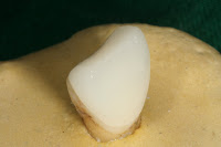
Ceramic veneers: Colour change in luting agents used for bonding them.

Ceramic Veneer
Tooth preparation

Veneer bonded to tooth surface


This work is ongoing.

I am working on the hypothesis that there is a change in the colour of a resin luting agent upon curing.

Resin based luting agents are used to bond ceramic veneers to teeth. It is well known that the thinness of the veneer will allow the shade of the luting agent to affect the over all shade of the final restoration.

So, basically I am making veneers of a standard thickness using E max (Ivoclar-Vivadent)on prepared, freshly extracted human central incisors.I will be bonding these veneers using 2 luting agents- Variolink II (dual cured) and Choice 2 (light cured). The colour of the tooth with the veneer placed on it will be assessed in terms of the L , a, b values using a spectrophotometer. This assessment will be done after placing the veneer without the luting agent and after that, before and after the curing of the luting agent.

The study should give us insights about the colour change in the tooth-veneer complex that we should expect after the veneer is bonded, using different types of luting agents.

The pilot study is done and we are quite surprised by the results as of now.

What interested me in this project?... Well, the chance of learning and prepping teeth for veneers, discovering how to make a porcelain veneers, the aspect of bonding and spectrophotometric testing... and above all- Its Prosthodontics!!

Medicon 2009


I was delighted to be given an oppurtunity to present my research at a National Conference dedidcated to research presentations only, that too in all disciplines of medicines, not just dentistry. This event is an annual event called Medicon


Medicon 2009 was an event where young scientists and future doctors met to present their research papers and share ideas in the fields of clinical and fundamental medicine, biomedicine and public health. A number of leading scientists and medical researchers gave encouraging and thought provoking talks to delegates.


I presented this poster which detailed the pilot study of my Project on Weathering of Silicone elastomers in Indian Climatic Conditions. Besides the poster and the research that I detailed in it, I also carried with me samples of Artificial prostheses such as Silicon Ears, Eyes and Noses. It was a great novelty for all the scientists and medical students there to see such prostheses for the first time!!. A lot of them felt the noses and ears in their hands and were amazed at the soft, skin-like texture of the silcione material used.

I would strongly urge you to take a look at the following organisation and sign up at the earliest.


INFORMER : informer.org.in

Medicine is a field constantly brimming with advances, the advances being investigative, diagnostic,therapeutic or otherwise. In a profession which demands a constant awareness of these advances, our aim is to consolidate and assess what a young medical researcher has to offer.

If India has to emerge as a global leader in medicine and health it is necessary that its medical research and education should be of global quality. Medical research continues to be a highly neglected area that should be strengthened urgently.


The need of a students' body to host the conference in various parts of the country in rotation and to promote research activities among the medical students at a national level was increasingly felt. It was decided that this should be an annual event finally converting it into a Medical Students' Science Congress in line with the Indian Science Congress. It was further recommended by the participants of the second conference that a National Body be founded. Subsequently a group of students under the able guidance of Dr Madhav G Deo worked out the Constitution of Indian Forum For Medical Students' Research (INFORMER) which is now a registered organization under the Societies Registration Act 1860 & B.P.T. Act 1950.

Objectives of INFORMER

1. To promote research and research-embedded education amongst undergraduate students both at the national and international level.
2. To secure and manage funds and endowments for the promotion of these objectives.
3. To perform all other acts that may assists in, or be necessary for the fulfillment of the above mentioned objectives as well as any additional objectives as approved by the Society.

I dashed down for a couple of days after finishing my exams. It was a joy to interact with so many students (mostly doing MBBS), staying at a hostel and above all, presenting my poster (thats the picture above) to the senior faculty who were experts from so many chosen fields!

A venture I would recommend highly!! I couldnt go for Medicon 2010 due to my final exams, but am keenly awaiting their next event and looking forward to enriching myself there. Cheers

Esthetic enhancements in Complete Dentures

I presented this paper at the National Dental Students Conference in Belgaum, 2009.

This work was published in the Journal Of the Indian Dental Association, JIDA, 2009, 3 ; 241- 243


Esthetics in complete dentures is more spoken of than practiced. Yet it remains a very important patient requirement in many situations and poses a challenge to the clinician.
Esthetics is defined as the skill and technique used to improve the art and symmetry of the teeth and face to enhance the appearance as well as the function of the teeth, oral cavity and face.1
The beauty about Esthetics lies in the fact that it is relative. What may be esthetically pleasing to an observer may not necessarily be the same to another. Inspite of this subjective nature of esthetics a well accepted fact is that an ideal conventional arrangement as is done for most complete denture cases gives dentures a standard/universal look which sometimes can look artificial.2
It is unreal to expect a natural look in elderly patients who wear dentures with nice shiny teeth, arranged in an ideal arch form and alignment, with perfectly sculpted gingival margins. A more realistic appearance for an older individual can be brought about by dentures which exhibit some attrition of teeth, gingival recession, tooth and gingival discolouration.

THE DENTOGENIC CONCEPT

The first attempt to incorporate esthetics as a separate important entity in complete dentures was by Frush and Fischer who introduced the Dentogenic concept. It is the art, practice and technique of creating an illusion of natural teeth in artificial dentures and is based on elementary factors suggested by the sex, personality and age of the patient.3
The 3 main aspects that can be looked at by the clinician to produce esthetically pleasing dentures are 3
1)Selection of the teeth4,5,6
2)Arrangement of teeth2
3)Characterization of denture


  • Teeth are selected according to their shape, size, colour and material to match the patients needs and requirements2. Dentogenics stresses that they should be selected keeping in mind the sex, personality and age of the patient .3
  • Smaller teeth with rounded line and point angles, rounded incisal edges give the teeth and the arrangement an over all feminine touch.
  • Larger teeth with flat edges, sharper line and point angles are more suited to a vigorous and masculine personality. 7
  • Chipping of teeth , overlapping of teeth are extremely common in natural dentitions and can be incorporated in the denture arrangements to make them look more natural. Some of the rules laid down in the literature for the above can be summarized as part of the ‘Dynesthetic concept’ of arrangement of teeth given by Frush and Fisher.8

Some of the principles highlighted in this concept can be used as ‘tips’ by the clinician as well as the laboratory technician to enhance the result and are summarized below:
  • Maxillary central incisors arranged in labioversion or with labial inclination look more pleasing.
  • Visibility of the teeth on speech must be such that the tip of the lateral incisor is just visible on speaking.
  • Central incisors must be arranged so that in young females and in young males they are 3mm and 2mm visible respectively below the lip in resting position. In older individuals the incisors are not kept visible at all or just at the level of the upper lip.8
  • The central incisors must dominate the anterior composition while the lateral incisors are considered to be the personality tooth. Sex determination comes from either rounding off the incisal edge of the lateral to depict feminity or squaring the edge to depict masculinity.9
  •  Diastemas must be judiciously placed so that they don’t serve as food traps and at the same time look esthetic2. All spaces must be V shaped to shed food. A diastema between the central incisors is avoided. Diastemas should be asymmetrically placed on either side of the dental arch.
  • The gum line must be placed at varying levels so that it is highest on the canines, followed by the central incisors and is the lowest on the laterals.2,8
  • The interdental papilla must be hygienic self cleansing and convex in all directions.

These rules are a guide and not a compulsion to be applied to all cases.
Characterization of dentures is the least explored of all options to add to the esthetic value of dentures2.It involves incorporation of subtle features in the dentures to give them some individuality.

The advantages of denture characterization are

a)It is a simple procedure
b)Does not require complicated/sophisticated/expensive materials or equipment.
c)It is not time consuming
d)It greatly enhances the esthetic outcome of the complete denture.

Characterization can be depicted easily by
a)Incorporation of stains.
b)Depth grinding.
c)Modification of incisal edges
d)Incorporation of restorations.
e)Incorporation of rotations.

Natural teeth and restorations like composites, usually tend to get stained by coffee, turmeric, paan, tobacco, and colouring agents in food. These stains are normally prominent on the proximal margins and cervical areas8.

Depth grinding involves sculpting of teeth to attain a desired morphology to compliment the patient. The mould of the tooth can also be modified to incorporate abrasion, erosion, masculinity or feminity. For example, incisal edges can be flattened to depict attrition or the mesial and distal line angles can be modified to alter the facial contour of the tooth. Chipping of teeth is usually seen in older teeth, especially in the incisors. Such replication of broken down teeth can create a pleasing natural look. 9,10

Restorations such as composites for the anteriors and silver amalgam for the posterior teeth can be judicially incorporated in the dentures. Such restorations serve a dual purpose. Besides adding to the natural look of the dentures, they also serve to distract the onlooker’s mind from the fact that the patient has an artificial dentition.

The authors recommend the following indigenous method for denture characterization along the above mentioned principles.
1. Staining of denture teeth- Inorganic rare earth pigments are used. These are dissolved in monomer and painted on the surface of the tooth as required. A variety of colours may be used. For example, red pigments can be used to depict paan stains, brown pigments subtly placed around the margins of restorations can suitably depict coffee/food stains. The stains are covered by a thin layer of Bonding agent which is light cured.11

2. Depth grinding- This can be incorporated with a universal wheel, cylindrical and tapered fissure diamond burs used with a micromotor hand piece. The labial surface of teeth can thus be sculpted to alter the contour. The incisal edges can be flattened, or they can be chipped away, the proximal contact areas can be broadened to depict age changes. The modified teeth are then polished before arrangement.9,10

3. Restorations- In posterior teeth, cavity cutting is done and amalgam restorations can be incorporated, especially on the occlusal surface of mandibular teeth. Composites restorations with a subtle colour mismatch can be incorporated on the anterior teeth to give them a natural appearance.

4. Denture base characterization – A natural appearance can also be incorporated in the ‘pink’ zone of the denture. Gingival inflammations are commonly depicted by rolled marginal gingivae. Inflammation can be depicted by incorporating a generalized swelling of the attached gingiva with a loss of stippling. This is done at the stage of waxing up of the dentures. The same inorganic pigments described above for staining of teeth can be used for pigmenting denture bases. Red pigments can be painted in a dilute form to depict inflammations while a mix of brown and black can be painted on in patches to simulate the melanin pigmentation of attached gingiva which is a very common Asian trait. These painted areas are sealed in by covering a thin layer of clear self cure acrylic resin which is then polished.


Therefore it may be realized that the esthetic outcome of complete dentures can be very easily enhanced by application of principles of dentogenics and carrying out denture teeth and denture base characterization wherever possible. Taking an extra step to pay due attention to the esthetics of a denture can go a long way towards ensuring success with complete denture treatment.
Characterization also is of great importance when the opposite dentition is a natural one especially that with spacings, stains  and pigmentation.


References

1)Mosbys dental dictionary,Elsevier,2nd edition,pg:229,2008
2)Charles M.Heattwell,Arthur O.Rahn,Heartwell ,4th edition-1984,Akshar Pratiroop Pvt.Ltd,pgs:97-104,309-324,375-389.
3)Frush J,Fisher R:Dentogenics:Its practical application.J.Prosthet Dent 1956,9:914-921,1959
4)Zarb,Bolender,Boucher,Elsevier,12th edition,pg:298-320.
5)Sheldon Winkler,Winkler,2nd edition 2000,A.T.B.S Publishers&Distributors, pg:202-216.
6)Felix.A.French,The selection and arrangement of the anterior teeth in Prosthetic dentures,1951
7)Frush J, Fisher R : How dentogenics interprets the personality factor. J Prosthet Dent 1956, 6: 441-449
8)Frush J, Fisher R : The dynesthetic interpretation of the dentogenic concept. J Prosthet Dent 1958, 8: 558-581
9)Frush J, Fisher R : How dentogenic restorations interpret the sex factor. J Prosthet Dent 1956, 6: 160-172
10)Frush J, Fisher R : The age factor in dentogenics. J Prosthet Dent 1957, 7: 5-13

Hardening of Maxillofacial Elastomers in Indian Climatic conditions.

Workers: Tania Sethi, Dr Mohit Kheur*, Dr Trevor Coward**, Colin Haylock***

* Professor, Prosthodontics, M.A Rangoonwala Dental College,Pune
** Head, Dept of Maxillofacial Prosthetics, Kings College, London,UK
*** Dept of Maxillofacial Prosthetics, Kings College, London,UK


Statement of Purpose

Maxillofacial prosthetics plays a key role in the over all rehabilitation of patients who have undergone extensive surgery following tumour resections and trauma. Silicone materials have over taken conventional acrylic resins and have become the materials of choice for the fabrication of such maxillofacial prostheses. This is mainly because of their clinical inertness, strength, durability, ease of manipulation and the comfort that they offer to patients, compared to the older materials. However such prostheses need to be periodically replaced. The main reason for replacement is the degradation of its colour and physical properties. The rate and amount of these changes can be variable depending on the climatic conditions and environment that the prosthesis is worn in. Such changes could also occur in varying proportions depending upon whether the material is cured at room temperature or by the application of heat in a hot air oven.
There is no report in the literature of the effect of the warmer, more humid Indian sub- continental environment on the rate of degradation of silicones using the above mentioned parameters and hence this study has been designed.

MATERIALS STUDIED
a) M 511 – 10: 1 platinum based system (Principality Medical, UK)
b) Z004 – 1:1 platinum based system (Principality Medical, UK)

TESTING APPARATUS


Hardness test – IRHD (Int Rubber Hardness test Device ) I have put a picture at the top.
FABRICATION OF THE SAMPLES

Sample size

A total of 60 samples were fabricated for each material.
30 samples were denoted as Group A and 30 samples as Group B.

Group A included 30 samples of each material made by room temperature curing.

Sub Group A1 – Samples of M511 by room temperature curing – Total 30 samples
Sub Group A2 – Samples of Z 004by room temperature curing – Total 30 samples

Group B included 30 samples of each material made by heat application.

Sub Group B1 – Samples of M511 by heat application – Total 30 samples
Sub Group B2 – Samples of Z 004 by heat application – Total 30 samples


Test and Control Group:

The samples are shown above.

Test group - 13 samples each from Sub Groups A1, A2 , B1, B2. – For the Patient study.

10 samples each from Sub groups A1 A2 B1, B2 – Weathered on roof top.

Control Group – 7 samples each – Sub Groups A1, A2 , B1, B2.


Manipulation of the material -

Each of the materials were handled in strict compliance with the manufacturers instructions. To achieve maximum consistency among specimens within the same Group, all such specimens were processed together.

The base and the catalyst was weighed using an electronic weighing scale and vacuum mixed.

The ratio used was 182 gm base and 18 gm of catalyst for the M 511 material.
The ratio for Z 004 was 100gm base and 100 gm of catalyst.

Each of the 200 gm of silicone were coloured using
Dry earth pigments (Cosmesil pigments, Technovent Ltd UK)

Umber-0.1gm
Dark yellow– 0.1 gm
Blue- 0.01 gm
Red-0.01gm

(This gave us a shade resembling Asian skin colours)

Thus from the same mix, the silicone was divided into 2 groups.


The Group A samples were left to cure at room temperature for 10 hours
The Group B samples were placed in a hot air oven at 85 deg Celsius for 1 hour.

STORAGE OF THE SAMPLES

7 samples that form the Control group were stored in an airtight container free from exposure to light and at ambient room temperature throughout the duration of the study.

10 samples in the study groups A and B of each material type were suspended from wooden racks by stainless steel suture material and placed on the roof of the Dental Institute.

13 samples each, from Group A and Group B were worn on the wrist of Dental students in the form of an attractive bracelet. Each bracelet had 1 sample each from Groups A1, A2, B1 B2. This was done to simulate the use of prosthesis on human skin.


TESTING PROCEDURE

Testing of the samples, (for both control group and the test groups) was done for
a) The colour change and
b) Change in hardness.

These changes were studied by repeated testing which was done at 3 stages –

Stage 1 - Immediately after curing. This time is to be used as the baseline time.
Stage 2 - 3 months from baseline.
Stage 3 - 9 months from baseline.


Hardness test – Hardness Was calculated by applying the IRHD at 3 points on each sample at every time interval mentioned above. The mean was taken as the mean hardness of each sample and the change was calculated at each of the intervals mentioned above.

(The IRHD is a very sensitive computerized hardness testing device that can measure hardness up to 2 decimal points. The previous studies using Shore A Durometers have measured hardness with certain limitations like limited sensitivity, less accuracy and are subject to human error while using the machine. The IRHD has a computer driven Shore A indenter)

The results thus were analyzed, subjected to a statistical analysis and appropriate conclusions were drawn.

My Results and Conclusions:
Overall, at 3-months the average hardness change in both the materials was significantly approximately similar.

However,
Material 2 Simweathering samples had smaller change compared to Material 1 samples.

In general, at 6-months the average hardness change in material 1 (M 511) was significantly lower compared to Material 2 (Z 004).
However,
Material 1 Cooled had smaller change compared to Material 2 cooled.

Material 1 Clinical and Control samples showed smaller change compared to Material 2 Clinical and Controls samples.

Overall, at 9-months there was no statistically significant difference in hardness change between the two materials.
However,
Material 1 Cooled had smaller change compared to Material 2 cooled.

Clinical and Control samples of Material 1 Showed significantly smaller hardness change compared to respective samples of Material 1.

Simweathiering sample of Material 2 Showed significantly smaller hardness change compare to Control sample of Material 1.


References:

1. Interactions of pigments and opacifiers on color stability of MDX4-4210/type A maxillofacial elastomers subjected to artificial aging
Sudarat Kiat-amnuay, Trakol Mekayarajjananonth, John M. Powers, Mark S. Chambers, James C. Lemon
J Prosthet Dent 2006 ;95, 3, 249-257

2. Ultraviolet radiation-induced color shifts occurring in oil-pigmented maxillofacial elastomers
Mark W. Beatty, Gordon K. Mahanna, Wenyi Jia
J Prosthet Dent 1999 ; 82, 4, 441-446

3. In vitro evaluation of color change in maxillofacial elastomer through the use of an ultraviolet light absorber and a hindered amine light stabilizer
Ngoc H. Tran, Mark Scarbecz, John J. Gary
J Prosthet Dent 2004 ;91,5, 483-490

4. Color stability of facial silicone prosthetic polymers after outdoor weathering
Gregory L. Polyzois
J Prosthet Dent 1999 ; 82, 4,447-450

5. Physical properties of a silicone prosthetic elastomer stored in simulated skin secretions
Gregory L. Polyzois, Petroula A. Tarantili, Mary J. Frangou, Andreas G. Andreopoulos
J Prosthet Dent 2000 ; 83, 5,572-577

6. Accelerated color change in a maxillofacial elastomer with and without pigmentation
John J. Gary, Eugene F. Huget, Larry D. Powell
J Prosthet Dent 2001 ; 85, 6, 614-620

7. Color stability and colorant effect on maxillofacial elastomers. Part III: Weathering effect on color
Steven P. Haug, Carl J. Andres, B. Keith Moore
J Prosthet Dent 1999 ; 81,4, 431-438

8. Color stability and colorant effect on maxillofacial elastomers. Part I: Colorant effect on physical properties
Steven P. Haug, Carl J. Andres, B. Keith Moore
J Prosthet Dent 1999 ; 81,4,418-422)

9. Color stability and colorant effect on maxillofacial elastomers. Part II: Weathering effect on physical properties
Steven P. Haug, B. Keith Moore, Carl J. Andres
J Prosthet Dent 1999 ; 81,4, 423-430

About me: Welcome to my blog..


Hi there. I am presently graduating from M. A Rangoonwala College of Dental Sciences and Research centre, Pune, India. I have a very keen interest in Prosthodontics and Restorative Dentistry.
I am very passionate about my work and this passion has enabled me to undertake a few short research projects in the area of Prosthodontics.
This blog is a show case of this work and I sincerely hope you like it. Your comments and views are most welcome!